Better Results for Skin Cancer Surgery
University of Rochester Prof. Michael Giacomelli recently received NIH* funding for his project, "Fluorescence microscopy for evaluation of Mohs surgical margins". His research objective is to enable real-time evaluation of pathology in skin tissue with an order of magnitude reduction in processing time as compared to frozen sections.
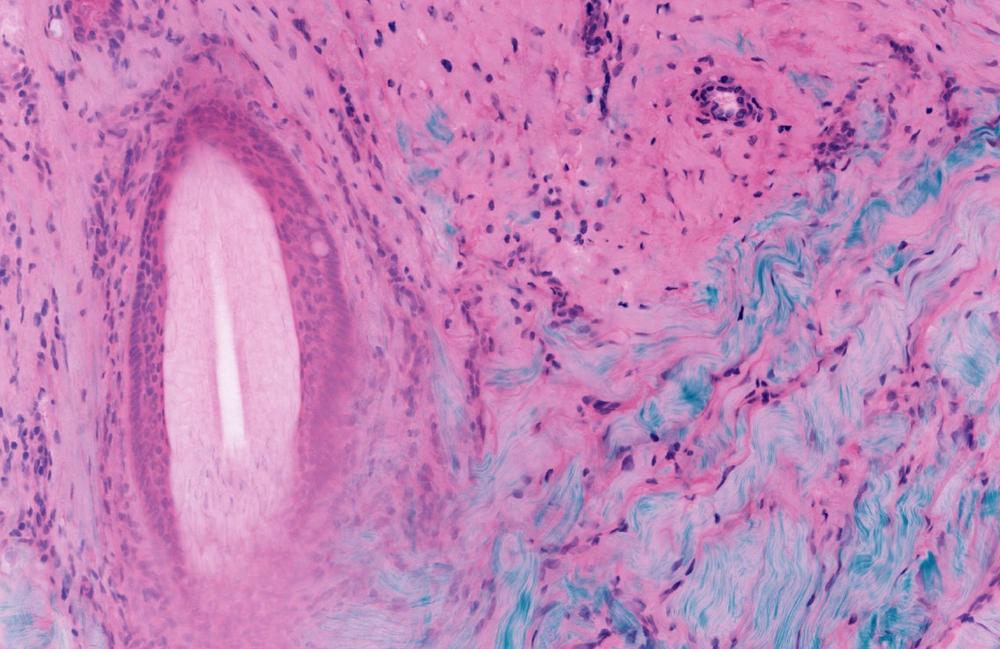
The most common forms of cancer worldwide are basal cell carcinoma and squamous cell carcinoma. And their incidence is showing a rapidly increasing trend in the last years. Mohs surgery is a widely used technique for the treatment of nonmelanoma skin cancer that obtains extremely low recurrence rates by imaging tissue as it is removed from the body to ensure complete resection. Mohs Micrographic Surgery, also called Mohs Surgery, is a specialized technique to remove non-melanoma skin cancers.
Fluorescence microscopy enables real-time evaluation of pathology
Nonmelanoma skin cancer (NMSC) is primarily diagnosed by histologic analysis of skin biopsy specimens in kerosene sections, which takes days to weeks before a formal diagnosis is made. Two-photon fluorescence microscopy (TPFM) has the potential for point-of-care diagnosis of NMSC and other dermatologic conditions, which could allow diagnosis and treatment at the same site.
Easy-to-use microscopes for clinics and doctors
Prof. Giacomelli developed a way to image multiple depth slices without frozen section processing – by implementing two-photon imaging, widely used in the neurosciences to noninvasively image slices at different depths in living brains. The lab team’s research has adapted these fluorescence imaging technologies with rapid tissue labeling and image processing technologies. This enables real-time assessment of pathology in skin tissue. processing time compared to frozen sections has been reduced by an order of magnitude. The data collection time is further reduced using the lower-noised silicon photomultiplier detectors developed by Giacomelli’s group.
Lasers enable clinical imaging of pathology in living human tissue
2-photon fluorescence microscopy can provide rapid point-of-care diagnosis of non-melanoma skin cancer through real-time imaging of fresh tissue biopsies.
The femtosecond laser system (FemtoFiber ultra 920) chosen for the next iteration of the Giacomelli lab’s microscope is a possible solution for applications in non-linear microscopy like two-photon excitation of fluorescent proteins and SHG based contrast mechanisms. With an emission wavelength of 920 nm, it provides the highest peak power for imaging with green and yellow fluorescent protein markers (GFP, YFP) commonly used in pathology, neurosciences and other laser-related biophotonic disciplines. Researchers appreciate the usability of the system since it is both, maintenance free and very compact.
About Giacomelli Lab / University of Rochester
The Giacomelli Lab focuses on the application of multiphoton and fluorescence imaging to surgical pathology and clinical medicine. The lab team designs custom multiphoton and fluorescence microscopes, algorithms, and electronics that enable surgical and clinical imaging of pathology in living human tissue with an emphasis on building instruments that can be directly used by clinicians and non-engineers.
* US National Institutes of Health (NIH) is the largest public funder of biomedical research in the world, investing more than $32 billion a year to enhance life, and reduce illness and disability? NIH funded research has led to breakthroughs and new treatments, helping people live longer, healthier lives, and building the research foundation that drives discovery.
TOPTICA has been developing and manufacturing high-end laser systems for scientific and industrial applications for more than 20 years. Our portfolio includes diode lasers, ultrafast fiber lasers, terahertz systems and optical frequency combs. TOPTICA today has 450 employees in 6 commercial entities with a consolidated group revenue of 100 Mio € (about 107 Mio $).
TOPTICA Photonics SE
Lochhamer Schlag 19
82166 Gräfelfing
Telefon: +49 (89) 85837-0
Telefax: +49 (89) 85837-200
https://www.toptica.com
![]()