Hamamatsu Photonics succeeded in creating a technology that upgrades the spatial resolution of two-photon excitation fluorescence microscopy
Applying results from this research will easily enhance the resolution of two-photon excitation microscopes equipped with an SLM. This new microscopy technique is being put to practical use across a broad range of fields including neuroscience and biology. It allows for detailed measurements of deep regions in biological samples and highly accurate observations of changes in the state of organelles that make up cells. This makes it a promising tool for applications in research on brain functions, kidney disease and other illnesses.
The results from this research were published on April 20 in the leading scientific journal on neuroscience “Frontiers in Neuroscience”. Results were achieved by a joint research effort with a laboratory endowed for Biophotonics Innovation and from a course on Virology and Parasitology offered at the Hamamatsu University School of Medicine.
How fluorescence microscopy works
*1: A spatial light modulator or SLM is an optical device that utilizes a liquid crystal to control the wavefront of an incident light such as from lasers to adjust the wavefront shape of the reflected light. Using a spatial light modulator allows freely controlling the laser beam pattern for example to branch the incident light and correct its distortion.
Research background
In scientific fields such as neuroscience, biology and medicine, it is necessary to clearly observe deeper positions of thick biological samples such as brain tissues. Two-photon excitation fluorescence microscopy utilizes near-infrared light which penetrates well into biological samples, reaching deeper positions compared to ordinary fluorescence microscopes using visible light. In deep regions of a sample, however, aberrations (*2) are likely to occur depending on the lens’ characteristics and the sample itself, causing a significant loss in resolution. To cope with this problem, two-photon excitation fluorescence microscopes equipped with an SLM are being designed for everyday use. They cancel out the aberrations by feeding a hologram pattern into the SLM.
It is the growing demand from universities and research institutes for higher resolution observation that prompted us to work jointly with the Hamamatsu University School of Medicine. Together, we created this technology improving the resolution of two-photon excitation fluorescence microscopy for practical applications.
How SLM works to correct aberrations
When the laser light is emitted on the SLM, it reaches a hologram pattern for aberration correction, which is fed from a computer. It is then reflected back with its aberrations corrected. In two-photon excitation fluorescence microscopy, the sample is irradiated with the laser light with its aberrations corrected, so specific areas provide a sharper and clearer image.
*2: Aberration is a distortion of an optical wavefront. The presence of aberrations causes loss of light condensing capability, lowers microscope resolution and laser processing efficiency, etc. Aberrations can be cancelled out by controlling the wavefront with an SLM.
Overview of research results
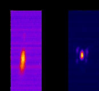
In this research, we carefully considered the number and shapes of hologram pattern rings based on our unique optical control technology and eventually succeeded in finding an optimal pattern that effectively improves the resolution. To further boost it, we also added an optical component that controls the light polarization (*3). By feeding the optimal hologram pattern and adding one optical component in this way, the resolution can be enhanced by about 20% without having to make drastic changes in the microscope optics design.
We will continue this R&D of improving resolution to widen the scope of practical uses and applications.
*3: Polarization is a condition in which light oscillates only on a certain plane.
Measurements of spherical fluorescent nanodiamonds at a depth of 50 μm from the sample surface.
Results from this research show a higher resolution not possible up until now.
Hamamatsu Photonics Deutschland GmbH
Arzberger Str. 10
82211 Herrsching am Ammersee
Telefon: +49 (8152) 375-0
Telefax: +49 (8152) 375-199
http://www.hamamatsu.com
![]()